What You Will Learn:
Section 1: History & Mycobiome
Part 1: The Name & Origins
Part 2: The Mycobiome & Natural Overgrowth
Section 2: Biology & Cellular Mechanisms
Part 1: Lipid Dependency & Sebum Breakdown
Part 2: How Malassezia Repairs the Skin Barrier
Part 3: How Follicular Breakouts Happen
Part 4: Bioactive Metabolites & Related Skin Conditions
Section 3: Triggers, Symptoms & Differential Diagnosis
Part 1: Demographics & Misdiagnosis
Part 2: Triggers
Part 3: Clinical Presentation
Part 4: Differential Diagnosis (Fungal Acne vs. Lookalikes)
Section 4: Treatment & Prevention
Part 1: Microbiome Balancing vs. Eradication
Part 2: Step-by-Step Treatment Plan (EADV Consensus)
Part 3: Active Ingredients
Part 4: Safety
Section 1: History & Mycobiome
Part 1: The Name & Origins
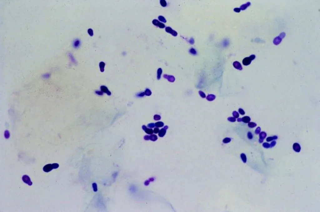
Popularly known as “fungal acne,” Malassezia (Pityrosporum) folliculitis (MF/PF) is not actually true acne. It is an inflammation and overgrowth of the hair follicle caused in over 90% of cases by Malassezia yeasts [2].
The Evolution of the Name: A yeast correlating with the pigment-altering skin condition tinea (pityriasis) versicolor (TV/PV) was first discovered in 1846 and named Malassezia furfur in 1853. In 1874, researcher Louis-Charles Malassez identified yeast-like cells within the stratum corneum, which were later cultured and named Pityrosporum ovale. Malassezia was designated as a distinct genus in 1889, and after decades of treating Pityrosporum and Malassezia as a separate genera, taxonomic revisions established Malassezia as the official genus name [4, 6].
The Discovery of “Fungal Acne”: Malassezia folliculitis was first documented in 1969 as an acne-like eruption triggered by broad-spectrum antibiotic use. It was officially recognized as a distinct clinical condition in 1973 [1].
Taxonomic Classification & Key Cutaneous Species: Malassezia is a monophyletic (sharing a single common ancestor) genus of lipophilic (oil-loving), dimorphic (able to exist in both a yeast spore phase and a filamentous hyphal phase) basidiomycetous yeasts (phylum Basidiomycota, class Malasseziomycetes) comprising 17-21 recognized and cultivated species. Ten species hold primary clinical importance on human skin: M. globosa, M. restricta, M. sympodialis, M. dermatis, M. japonica, M. obtusa, M. slooffiae, M. furfur, M. pachydermatis, and M. nana, alongside newly cultivated species such as M. arunalokei [5, 7].
Summary: “Fungal acne” is not true acne, but an overgrowth of oil-loving Malassezia yeast (formerly called Pityrosporum) inside the hair follicle that was first linked to antibiotic use in 1969 and recognized as a distinct condition in 1973. Out of 17-21 known Malassezia species, 10 play a primary role on human skin.
Part 2: The Mycobiome & Natural Overgrowth
Dominance in the Healthy Skin Microbiome: Rather than being a foreign invader, Malassezia is a normal commensal organism (a resident microbe that normally lives harmlessly on the body) that makes up greater than 80% of the total fungal population (mycobiome) of human skin [6] and is detected in the stratum corneum of 75%-98% of healthy adults [1]. Colonization begins immediately after birth, detected in 89% of newborns on day 0 and 100% on day 1 [3], and ubiquitously colonizes infants by 3-6 months of age [6]. Because this yeast depends on host skin oils (lipids), it concentrates in oil-rich (seborrheic) zones such as the face, scalp, and thorax, though it inhabits almost all body sites except the feet [5].
Predominant Species: M. restricta and M. globosa are the most abundant species on both healthy and diseased human skin, followed by M. sympodialis and M. furfur [6]. Although M. furfur is less prevalent on healthy skin, its abundance rises significantly in atopic dermatitis (eczema) and seborrheic dermatitis [7]. Multiple species frequently cohabit the exact same skin site simultaneously, most commonly in pairs of M. globosa-M. restricta, M. globosa-M.sympodialis, and M. restricta-M.sympodialis [8].
Why It Is Not Contagious: Malassezia folliculitis is benign and not contagious. While different species may be isolated from breakouts, all Malassezia species produce the exact same clinical presentation. In a clinical study by Durdu et al. (n = 49 MF patients), rDNA analysis identified M. globosa (69.4%) as the most common species inside active lesions, followed by M. sympodialis, M. restricta, and M. furfur. In 72% of cases, the exact same species were found on both breakout-affected (lesional) and clear (non-lesional) skin of the same patient [1]. Similarly, a cross-sectional PCR and culture study (n = 32 MF patients vs. n = 40 healthy controls) found the exact same predominant species across all groups with no significant difference in species count or total fungal burden: MF lesions had 1.7±0.7 species, non-lesional patient skin had 1.5±0.9 species, and healthy control skin had 1.9±0.8 species (p > 0.05) [3]. This proves that “fungal acne” is caused by an internal overgrowth or dysregulation of a person’s existing resident flora, not an outside infection.
Summary: Malassezia is a normal, harmless resident microbe that makes up over 80% of our skin’s fungal population from birth, with M. restricta and M. globosa being the most common species. Because the exact same yeast species are found on both clear and breakout-prone skin, “fungal acne” is not contagious, it is simply an internal overgrowth of your skin’s own resident flora.
Section 2: Biology & Cellular Mechanisms
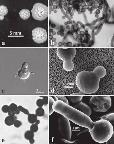
At the microscopic level, Malassezia is a polymorphic (taking multiple shapes), lipophilic, monopolar budding yeast (daughter cells bud repeatedly from a single site at one end of the mother cell) with a thick, multilayered cell wall. Specifically, M. globosa has spherical/globose cells, while M. sympodial exhibits sympodial budding [1]. Within human skin, Malassezia interacts both directly and via chemical messengers (mediators) with keratinocytes (epidermal skin cells); Langerhans cells and dendritic cells (antigen-presenting immune cells); melanocytes (pigment-producing cells); mast cells (histamine-releasing cells); T cells; B cells; and both the innate and acquired immune systems [5].
Part 1: Lipid Dependency & Sebum Breakdown
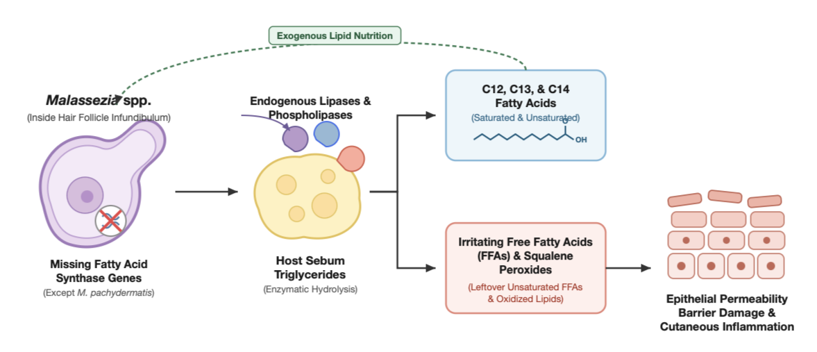
Overview: Because human Malassezia species lack the gene required to make their own fatty acids, they must feed on your skin’s sebum. To digest your sebum, the yeast releases digestive enzymes that split sebum triglycerides apart, leaving behind components that damage the skin barrier and trigger inflammation.
Part 1a: Diagram Breakdown
Missing Enzyme Genes: With the sole exception of M. pachydermatis, Malassezia species lack fatty acid synthase genes. Because they cannot manufacture their own fatty acids, they are strictly dependent on exogenous (externally supplies) host lipids to survive [6].
Carbon Chain Requirements: Specifically, Malassezia species require C12, C13, and C14 saturated and unsaturated fatty acids [1].
Enzymatic Sebum Breakdown (Hydrolysis): Malassezia secretes endogenous lipases and phospholipases (digestive enzymes) to hydrolyze (chemically split apart) host sebum triglycerides into free fatty acids (FFAs) so the yeast can feed [1]. However, this breakdown also generates irritating leftover FFAs and squalene peroxides (oxidized squalene), which can damage the epithelial permeability barrier and trigger cutaneous inflammation [5].
Part 2: How Malassezia Repairs the Skin Barrier
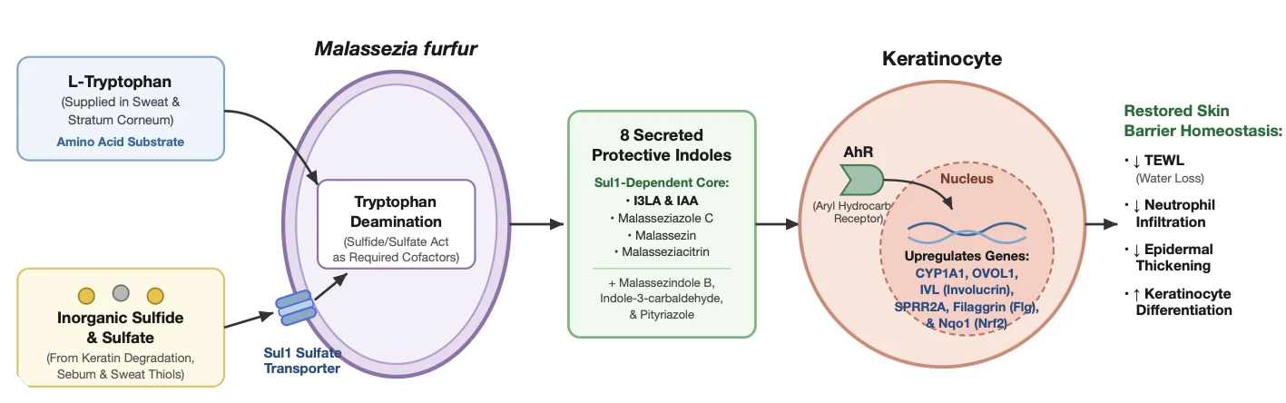
Overview: Malassezia actively protects and repairs your skin barrier when balanced. M. furfur produces protective compounds, turning on barrier-repair genes to lower transepdiermal water loss (TEWL) and calm inflammation.
Part 2a: Diagram Breakdown
Tryptophan & Sulfur (Sul1) Requirements: M. furfur metabolizes the amino acid L-tryptophan (naturally supplied in sweat and the stratum corneum) into 8 specific AhR-activating indoles: indole-3-lactic acid (I3LA), indole-3-acetic acid (IAA), malasseziazole C, malassezindole B, indole-3-carbaldehyde, pityriazole, malassezin, and malasseziacitrin. To synthesize its core protective indoles (I3LA, IAA, malasseziazole C, malassezin, malasseziacitrin), M. furfur requires its Sul1 sulfate transporter to take up inorganic sulfide and sulfate (naturally supplies on skin via keratin degradation, sebum, and sweat thiols), which act as required cofactors for tryptophan deamination [7].
Barrier Gene Upregulation & Anti-Inflammation: When M. furfur produces these indoles, they bind AhR keratinocytes and upregulate critical barrier-building and antioxidant genes: CYP1A1, involucrin (IVL), small proline-rich protein 2A (SPRR2A), OVOL1 (a keratinocyte differentiation regulator), filaggrin (Flg), and Nqo1 (part of the Nrf2 antioxidant pathway). In vivo, indole-producing M. furfur (or topical application of I3LA and IAA) reduces abnormal skin/epidermal thickening, blocks inflammatory neutrophil (white blood cell) infiltration, and lowers transepidermal water loss (TEWL) in barrier-damaged skin. Conversely, if M. furfur is deprived of tryptophan or sulfur uptake, it fails to produce protective indoles and instead worsens inflammation and TEWL [7].
Part 3: How Follicular Breakouts Happen
Overview: When pores become occluded or the microbiome is disrupted, Malassezia multiplies deep inside the follicle. Its lipid-digesting enzymes and cell wall components release inflammatory cytokines that swell and rupture the pore wall, causing uniform, itchy red papules and pustules.
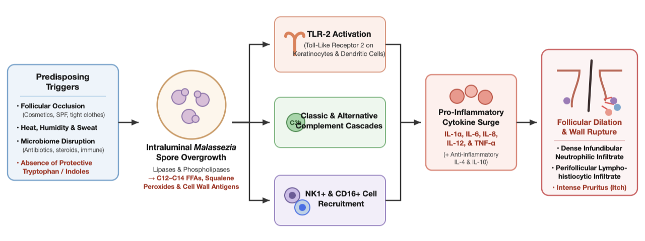
Part 3a: Diagram Breakdown
Receptor & Immune Activation: When pore occlusion, heat, humidity, sweat, antibiotic flora disruption, or immunosuppression occur, alongside an absence of protective tryptophan/indole signaling, Malassezia proliferates deep inside the follicle [3, 7]. Its lipase-generated C12-C14 free fatty acids, squalene peroxides, and fungal cell wall components activate TLR-2 on keratinocytes and dendritic cells, trigger both the classic and alternative complement cascades (innate immune proteins that amplify inflammation), and recruit NK1+ and CD16+ immune cells [5].
Cytokine Release & Follicular Wall Rupture: This immune activation releases pro-inflammatory chemical messengers (cytokines) including IL-1α, IL-6, IL-8, IL-12, and TNF-α (alongside anti-inflammatory IL-4 and IL-10), which damage the follicular barrier and cause the follicle to swell [5]. When the dilated follicular wall ruptures, it triggers an infundibular neutrophilic (pus-forming white blood cells inside the upper pore) and perifollicular lymphohistiocytic infiltrate (immune cells surrounding the pore), resulting in classic pustules, papules, and intense pruritus (itching) [1].
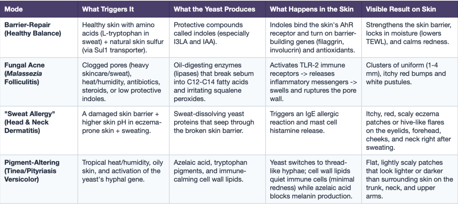
Table 1: How Malassezia Acts on Skin - Protective vs. Disease Modes
Part 4: Bioactive Metabolites & Related Skin Conditions
Malassezia species oxidize unsaturated fatty acids into dicarboxylic acids (specifically azelaic acid) which competitively inhibit the pigment enzyme tyrosinase and exert direct cytotoxic effects on hyperactive melanocytes. This explains why Malassezia causes pale, depigmented patches (hypochromia) in tinea (pityriasis) versicolor (TV/PV). Additionally, in TV, pathogenic hyphal growth is linked to the activation of the M. globosa MGL_3741 gene, which encodes the enzyme dihydroxy acid dehydrates (DHAD), an enzyme active in lesional skin but absent in healthy, non-lesional skin [5].
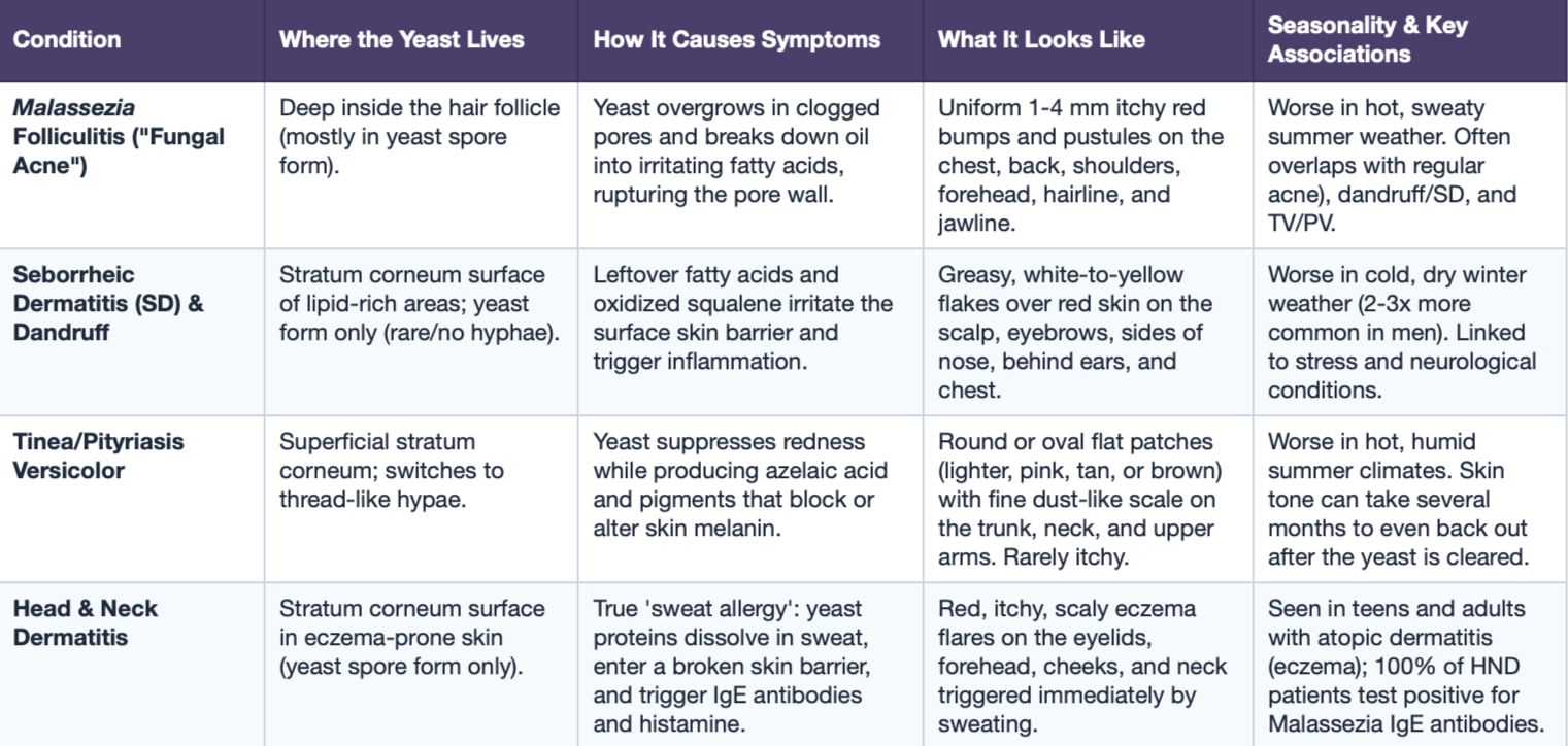
Table 2: The Four Malassezia Skin Conditions Side-by-Side
Because 18-40% of Malassezia folliculitis patients concurrently have Seborrheic Dermatitis and 6-16% have Pityriasis Versicolor [2], comparing how the same yeast behaves across these four conditions is essential for accurate differential assessment.
Section 3: Triggers, Symptoms & Differential Diagnosis
Part 1: Demographics & Misdiagnosis
Age & Sex: In a pooled PRISMA meta-analysis of N = 1,238 patients across 15 studies, Malassezia folliculitis predominantly affects males (64.0%, N = 970) and adolescents/young adults aged 11-30 years, with a pooled mean of 24.26/24.3 years (N = 403) [2]. This demographic skew aligns with peak androgen-driven sebum production in young adult males.
Multi-Year Diagnosis Delay: Because MF is so frequently misdiagnosed as standard acne vulgarism (AV), the average duration of disease prior to receiving a correct diagnosis in patients ranges from 10.9 months up to 61 months (over 5 years) [3].
Summary: Malassezia folliculitis most commonly affects teens and young adults aged 11-30 (with a 64% male skew due to higher androgen-driven oil production). Because it looks so similar to regular acne, patients go an average of 11 months to over 5 years before getting an accurate diagnosis.
Part 2: Triggers
Heat, Humidity & Summer Seasonality: High temperatures and humidity trigger the yeast’s switch to its pathogenic phase while increasing sweat and sebum production. In Lim et al. (n = 48), 83% of patients worked in a warm environment and 71% (n = 34/48) cited heat and sweating as direct aggravating factors [1, 3].
Follicular Occlusion (Cosmetics & Clothing): Tight, non-breathable (occlusive) clothing and heavy topical skincare products trap sebum, physically block (occlude) the follicular opening, and promote flares [1].
Oral & Topical Antibiotics: Across 10 pooled studies (N = 802), 40.5% of MF patients had a history of failed treatments, reaching 65-89.6% in individual cohorts [2]. First-line antibiotics (specifically tetracyclines like minocycline and doxycycline; macrocodes; and topical or oral clindamycin) wipe out normal commensal skin bacteria, destroying natural bacterial-fungal competition and allowing Malassezia to overgrow [3].
Oral & Topical Corticosteroids: Oral and topical steroids predispose hair follicles to Malassezia overgrowth. In a study by Yu et al,, 66-76.5% (n = 26/34) of patients clinically diagnosed with “steroid acne” actually had abundant Malassezia (P. ovale) spores inside their hair follicles [2].
Metabolic & Endocrine Conditions: Diabetes mellitus, Polycystic Ovarian Disease (PCOD/PCOS/PMOS), and hypothyroidism (elevated TSH) alter sebum and follicular homeostasis, predisposing individuals to MF [4].
Immunosuppresion: In a systemic review of 50 immunosuppressed MF patients, 94.1% developed MF after starting immunosuppressive therapy or becoming immunocompromised [3].
Summary: Flares are triggered externally by hot, humid summer weather, heavy sweating, tight clothing, and occlusive cosmetics. Internally, the biggest triggers are oral and topical antibiotics (which wipe out competing skin bacteria), corticosteroids (which account for up to 76.5% of “steroid acne” cases), hormonal conditions (diabetes, PCOD/PCOS/PMOS, hypothryoidism), and immunosuppression.
Part 3: Clinical Presentation
Classic vs. Atypical Lesion Morphology: Classic MF presents as monomorphic (uniform in size and shape), 1-2 mm (up to 2-4 mm) erythematous (red) or skin-colored follicular papules (solid bumps) and pustules (pus-filled bumps) centered directly around hair follicles [3].
Non-Comedonal: A defining clinical clue of MF is the complete absence of open or closed comedones (blackheads and whiteheads), deep nodules, or cysts (unless the patient has coexisting acne vulgars) [1].
Symptoms: Pruritus (itching) occurs in ~71.7-80% (pooled N = 540) of patients, with some patients also experiencing a slight burning sensation [1].
Location: Roughly 71.4% of patients have lesions across multiple seborrheic regions at the same time. Pooled PRISMA data (N = 1,238) ranks the chest (70.0%) and back/shoulders/upper arms (69.2%) highest, followed by the forehead/face (30.9%). [2].
Skin of Color Presentation: In darker skin, post-inflammatory hyperpigmentation (PIH) frequently masks underlying erythema (redness), causing MF to be misdiagnosed as follicular eczema, acne vulgars, or keratosis pilaris. Clinicians and estheticians should prioritize itching, monomorphic papules/pustules in seborrheic zones, the absence of comedones (unless patient has coexisting acne vulgars), and cross-polarized or UV dermoscopy [3].
Coexising Conditions: MF frequently overlaps with other seborrheic disorders, requiring multiple treatment strategies:
Acne Vulgaris (AV): Coexists in 12-27%+ of cases; Malassezia species are associated with the development of non-inflammatory acne vulgars [2].
Seborrheic Dermatitis (SD): Coexists in 18-40% of MF patients [2].
Tinea/Pityriasis Versicolor (TV/PV): Coexists in 6-16% of MF patients [2].
Summary: Classic Malassezia folliculitis looks like clusters or uniform (1-4 mm) red or skin-colored bumps and pustules with zero blackheads or whiteheads (unless patient has coexisting acne vulgaris), accompanied by itching in up to 80% of cases. It appears most often on the chest, back, shoulders, upper arms, forehead, hairline, and jawline, frequently coexisting alongside true acne vulgars (12-27%+) or seborrheic deramtitis (18-40%).
Part 4: Differential Diagnosis (Fungal Acne vs. Lookalikes)
Because Malassezia folliculitis is frequently confused with other conditions, clinicians and skincare professional must differentiate it from mimicking conditions including follicular eczema, keratosis pilaris (KP), and other similarly-presenting conditions.
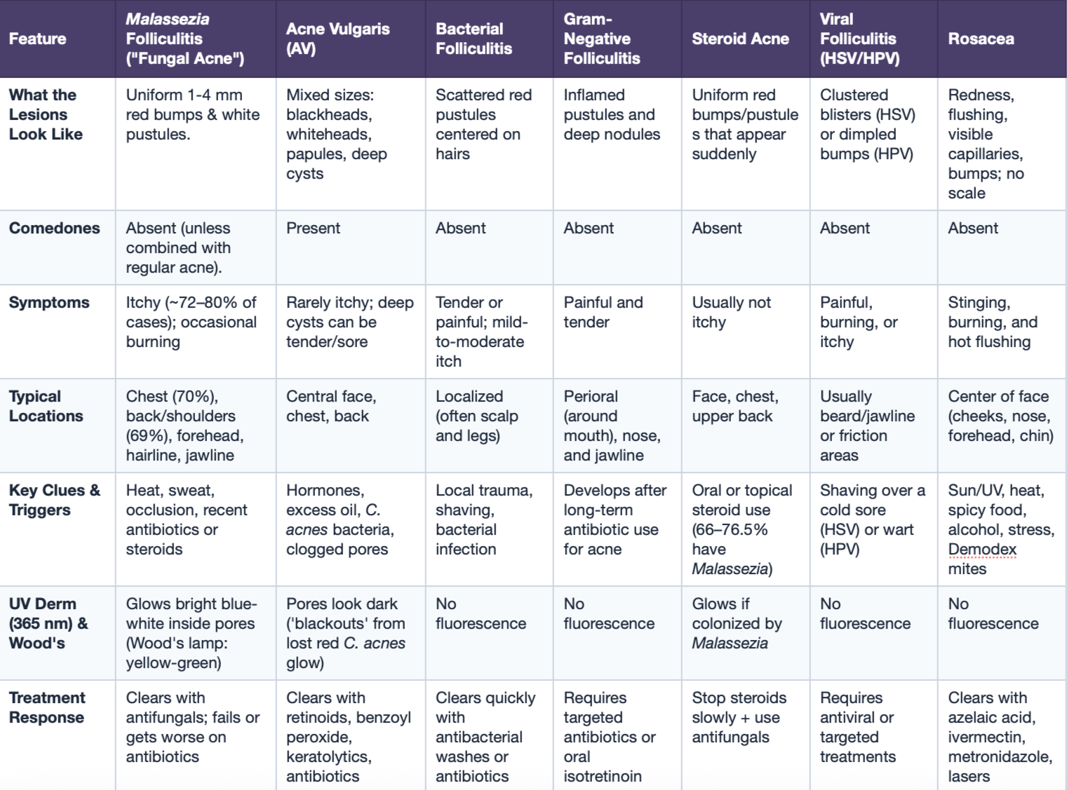
Table 3: Malassezia Folliculitis vs. Mimicking Conditions
Section 4: Treatment & Prevention
Part 1: Microbiome Balancing vs. Eradication
Because Malassezia is an essential resident of the healthy skin microbiome that actively repairs the skin barrier via AhR-activating indoles, the goal of treatment is not to permanently eradicate the yeast from the body, but to reduce follicular overgrowth and restore balance.
In a 6-month randomized comparative trial evaluating low-dose oral isotretinoin against a topical antifungal/keratolytic shampoo (piroctone olamine/LHA/salicylic acid), neither treatment eradicated Malassezia from the skin (93.5% positive cultures at baseline vs. 95.1% at Month 6), yet both treatments equally resolved clinical symptoms. Symptom resolution occurred via a microbiome shift: M. globosa and M. sympodialis decreased, while adapted M. restricta increased [8].
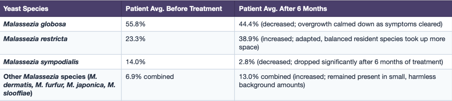
Table 4: How Treatments Balanced, Rather Than Eradicated, The Yeasts
Part 2: Step-by-Step Treatment Plan (2023 EADV Consensus)
First-Line for Healthy Patients (Topicals): Start with topical antifungals and keratolytics (specifically topical azoles, ciclopirox, propylene glycol, zinc pyrithione, piroctone olamine, selenium disulfide, or sulfur) for a 2-4 week trial. Topical therapy avoids systemic liver toxicity and achieves an 81.6% pooled improvement rate (reaching 100% in multiple cohorts), though it works roughly half as fast as oral pills (27±16 days for 2% ketaconazole cream vs. 14±4 days for oral itraconazole to flatten papules) [3].
First-Line for Severe, Resistant, or Immunocompromised Patients (Systemic Orals): A dermatologist may recommend oral systemic antifungals (itraconazole or fluconazole), which penetrates deep into the hair follicle and achieve a 92.0% pooled improvement rate (and 88.9-92.3% in immunosuppressed cohorts) [3].
Maintenance & Preventing Relapse: Relapse is extremely common once treatment stops. Patients must transition to intermittent maintenance therapy such as applying ketoconazole, ciclopirox, or selenium sulfide shampoo 1-2 times per week indefinitely, 50% propylene glycol intermittently, or monthly oral antifungals alongside modifying sweat and occlusive triggers [3].
Part 3: Active Ingredients
Disclaimer: This is not medical advice. Please consult a medical professional before making any medical decisions.
Oral Systemic Antifungals (Triazoles & Imidazoles)
Itraconazole (Evidence Level Ib-IIb): Dosed at 100-200 mg/day for 1-4 weeks (commonly 200 mg/day for 7-14 days) achieves 69-100% clinical recovery, 86.4% negative mycology at Week 5 (vs. 7.7% placebo), and flattens papules in 14±4 days [3].
Fluconazole (Evidence Level III): Dosed at 100-200 mg/day for 1-4 weeks (or 100 mg daily tapered to every other day) ahieves 80-100% clinical improvement including drastic improvement within 24 hours when paired with topical clotrimazole and tretinoin; it is very active against M. sympodialis and M. sloofiae, though inactive in vitro against M. globosa and M. restricta [5].
Oral Ketoconazole (Evidence Level IIb-III): Dosed at 200 mg/day (or pulsed over 8 weeks), achieving 75-92.1% clinical improvement alone and 100% clinical improvement when combined with topical 2% ketoconazole [3].
Topical Antifungals (Keotconazole, Ciclopirox & Terbinafine)
Ketoconazole 2% Cream or Shampoo (Evidence Level III/IV): Cream is applied twice daily for 4-6 weeks; shampoo is used daily during active breakouts, then 1-2 times per week for maintenance. Achieves 100% improvement with 2% cream twice daily in 27±16 days, 100% improvement combining 2% shampoo + cream, and 75% 6-month relapse prevention as a maintenance shampoo; it is more active against M. furfur than econazole or miconazole [1].
Ciclopirox Olamine 1% & Terbinafine 1% Cream (Evidence Level IV): Ciclopirox olamine 1% provides combined antifungal, keratolytic, and anti-inflammatory properties and is an EADV first-line and maintenance recommendation. Terbinafine 1% cream is effective topically, though oral terbinafine is ineffective for TV/PV and superficial Malassezia [5].
Keratolytic & Anti-Seborrheic Topicals
Selenium Disulfide 2-2.5% Shampoo/Lotion (Evidence Level IIb): Applied once weekly for 3-8 weeks (or daily for 3 days then weekly; lathered in shower for 3-4 minutes or left overnight) achieves 80% recovery at 4 weeks and 88% at 8 weeks (n = 22/25), combining antifungal and keratolytic actions to dissolve follicular plugs as an EADV first-line and top maintenance choice [3].
50% Propylene Glycol in Water (Evidence Level IIb): Applied twice daily for 3-8 weeks, achieving 75% recovery at 4 weeks and 100% recovery at 8 weeks (n = 12/12); acts as a dual keratolytic and antimicrobial EADV first-line and intermittent long-term suppressive therapy [3].
Zinc Pyrithione, Sulfur, Salicylic Acid, Resorcinol, Lithium Succinate: Zinc pyrithione and sulfur provide antifungal, keratolytic, and anti-inflammatory effects (EADV first-line) [3, 6]. Salicylic acid and resorcinol aid keratolysis (achieving 46% improvement with isoconazole) [1, 2, 6], while lithium succinate (in SD) blocks the availability of prerequisite lipids needed for Malassezia growth [5].
Dual Target Acne & MF Agents (Retinoids & BPO)
Topical Retinoids (Tretinoin) & Benzoyl Peroxide (BPO): Ideal when MF coexists with AV. Tretinoin provides anti-inflammatory and keratolytic benefits that normalized follicular desquamation, while BPO and salicylic acid aid keratolysis [1].
Oral Isotretinoin (Evidence Level III in MF; Level Ib in SD): Dosed at 0.65-0.85 mg/kg/day for 4-5 months for MF or 10 mg every other day for 6 months for SD. It shrinks sebaceous glands, reduces sebum synthesis, normalizes keratinization, and modulates TLR-2 and TLR-4 [8].
Part 4: Safety
Oral antifungals can cause nausea, vomiting, diarrhea, abdominal pain, and liver toxicity, and can affect liver, kidney, and hematopoietic functioning. They are strictly contraindicated in patients with active liver disease, elevated liver enzymes, prior antifungal hepatoxicity, hepatic drug-drug interactions, or women who are pregnant or trying to conceive (due to teratogenic birth-defect potential), and require periodic blood testing during prolonged courses [6].
Comment down below any thoughts, contributions, or requests for future posts! :)
References:
Rubenstein RM, Malerich SA. Malassezia (pityrosporum) folliculitis. J Clin Aesthet Dermatol. 2014 Mar;7(3):37-41. PMID: 24688625; PMCID: PMC3970831.
Green M, Feschuk AM, Kashetsky N, Maibach HI. Clinical characteristics and treatment outcomes of Pityrosporum folliculitis in immunocompetent patients. Arch Dermatol Res. 2023 Aug;315(6):1497-1509. doi: 10.1007/s00403-022-02506-0. Epub 2022 Dec 14. PMID: 36517586; PMCID: PMC9750048.
Martínez-Ortega JI, Mut Quej JE, Franco González S. Malassezia Folliculitis: Pathogenesis and Diagnostic Challenges. Cureus. 2024 Nov 11;16(11):e73429. doi: 10.7759/cureus.73429. PMID: 39664138; PMCID: PMC11633069.
Malgotra V, Singh H. Malassezia (Pityrosporum) Folliculitis Masquerading As Recalcitrant Acne. Cureus. 2021 Feb 24;13(2):e13534. doi: 10.7759/cureus.13534.
Saunte DML, Gaitanis G, Hay RJ. Malassezia-Associated Skin Diseases, the Use of Diagnostics and Treatment. Front Cell Infect Microbiol. 2020 Mar 20;10:112. doi: 10.3389/fcimb.2020.00112. PMID: 32266163; PMCID: PMC7098993.
Vest BE, Krauland K. Malassezia Furfur. [Updated 2023 May 22]. In: StatPearls [Internet]. Treasure Island (FL): StatPearls Publishing; 2023 Jan-. Available from: https://www.ncbi.nlm.nih.gov/books/NBK553091/.
Gushiken-Ibañez E, Stokmaier M, Barone G, Staropoli A, Karakaya T, Beer HD, Vinale F, Ianiri G, LeibundGut-Landmann S. Commensal yeast Malassezia produces tryptophan metabolites to promote tissue homeostasis via the aryl hydrocarbon receptor in mice. Nat Microbiol. 2026 Jul;11(7):1934-1948. doi: 10.1038/s41564-026-02382-8. Epub 2026 Jun 19. PMID: 42321537; PMCID: PMC13323057.
Kamamoto CSL, Nishikaku AS, Gompertz OF, Melo AS, Hassun KM, Bagatin E. Cutaneous fungal microbiome: Malassezia yeasts in seborrheic dermatitis scalp in a randomized, comparative and therapeutic trial. Dermatoendocrinol.2017 Oct 23;9(1):e1361573. doi: 10.1080/19381980.2017.1361573. PMID: 29484095; PMCID: PMC5821162.